
Beaumont Imaging Services
Cutting-Edge Technology to Give You Better Information
Baptist Hospitals of Southeast Texas uses some of the latest inpatient and outpatient imaging equipment available in the Beaumont, Texas area. We have partnered with a team of specially-trained and experienced technologists to benefit patients through innovative technology and the highest quality of patient care. We offer imaging services for all ages, from pediatrics to geriatrics.
With our commitment to diagnosis and prevention, we provide screenings for a variety of conditions including:
- Breast cancer
- Lung cancer
- LEAP(Lower Extremity Amputation Prevention)
- Cardiovascular disease
The Imaging Department supports diagnosis and treatment of Beaumont patients by providing:
- Diagnostic X-rays to provide images of a specific area
- CT scans to provide multiple views of an area or organ
- MRI imaging
- Ultrasound images with the SOMATOM Definition AS, to screen for a wide range of medical conditions from trauma to cancer
- Interventional radiologic imaging to target the treatment of some diseases
- 3-D and 4-D ultrasound imaging commonly used for fetal screening
- Digital mammography for early detection of breast cancer
- Cardiac calcium scoring to screen for heart disease
- Low-dose CT scans to check for early signs of lung cancer
- Vein Center to provide minimally invasive treatment for Varicose Veins and Venus Insufficiency
- SPG block to treat headache pain
List of Services
- CT Scans
- Special Procedures
- DEXA Scan
- Fluoroscopy
- Mammography
- MRI
- Nuclear Medicine
- PET/CT
- Ultrasound
- X-Ray
For more information about diagnostic imaging services, please follow the link to the Digisonics, Inc. website www.digison.net.
Special Procedures
The Radiology department at Baptist Hospital in Beaumont is committed to helping our patients find the right healthcare solutions for their needs. Our interventional radiologists specialize in small-area treatments such as catheter insertion, varicose vein therapy, and other procedures by using imaging equipment.
Interventional radiology is a medical specialty that performs various minimally-invasive procedures using medical imaging guidance, such as x-ray fluoroscopy, computed tomography, magnetic resonance imaging, or ultrasound.
Peripheral vascular disease (PVD), also known as peripheral artery disease (PAD), is a progressive disorder that occurs when blood flow to a body part outside of the brain or heart is reduced. This can happen when blood vessels narrow, become blocked, or spasm. The most common cause of PVD is atherosclerosis, which is a buildup of fatty deposits, or plaque, that can narrow arteries or veins.
Our Radiology department uses:
- X-ray
- Ultrasound
- Magnetic resonance imaging (MRI)
- Positron emission tomography (PET)
- Computed tomography (CT)
Treatments Near You
Our PET/CT scan equipment is the only of its kind in Southeast Texas. With the right radiology assistance, it’s possible to treat a number of conditions including headache pain.
Learn more about our Headache Treatment Center by clicking here.
To learn more about Varicose Vein Treatments, click here.
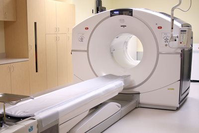
CT Scans
Providing Accurate Imaging Services at Baptist Hospital - Beaumont, Baptist Hospital - Orange, Baptist Outpatient Center
Using a rotating x-ray machine, a computed tomography (CT) scan provides a detailed image of the inside of the body in under a minute. At Baptist Hospitals of Southeast Texas, we use a 128-slice CT scanner that completes its scan in roughly 20 seconds or less. There is no more convenient way to obtain a comprehensive view of what goes on inside your body.
Preparing for Your CT Scan
Depending on what your scan procedure is being used to detect, you may be asked to ingest a dye called contrast that will help the scanner focus and identify circulation routes in your body. If this is the case, you should not eat or drink anything 4-6 before your procedure. Contrast may also be administered in the rectum with an enema or intravenously (IV), in which case you may feel slight discomfort as it enters the bloodstream. This sensation quickly subsides.
You will be required to wear a hospital gown and to remove all metallic jewelry as these objects will interfere with the scan procedure. Please read the American College of Radiology’s patient notice by clicking here.
DEXA Scans in Beaumont
High-Accuracy Imaging to Make Better Diagnoses at our Dauphin Women's Center and Lumberton Outpatient Center
While there are many scans available to check for vascular function and tissue health, bones require a specific scan using what is known as Dual Energy X-ray Absorptiometry (DEXA). As people grow older, their bones naturally lose mineral density and become more brittle. To better understand how to move forward with treatments and medication, Baptist Hospitals of Southeast Texas offers DEXA scans to help patients understand how they can best preserve their health.
What Is a DEXA Scan?
These imaging techniques pass a small amount of radiation through bones in the spine and in the hip and record how much is able to travel through bone and soft tissue and make it to the other side. By comparing different measurements, radiologists can determine how much mineral density a patient’s bones contain as well as how quickly the density is depleting. Measurements can be as sensitive as 2% different.
Why Undergo a DEXA Scan?
Bone diseases like osteoporosis do not appear right away. While a person may experience its symptoms, having a DEXA scan performed can help a person know exactly how healthy their bones are and make choices accordingly.
Scans can be used to:
- Detect the bone disease osteoporosis
- Identify risk factors that may later turn into osteoporosis
- Make decisions about hormone treatments following menopause
Preparing for a DEXA Scan
While these scans are usually very simple and do not require much preparation, it is important that you wear clothing that is free of metal, including jewelry, buttons, and buckles. During the scan, you will lie down on a table and have both your hip and your spine scanned at the same time. Unless you suffer from back pain of some sort, lying on the table for the scan will not provide any unusual discomfort.
Fluoroscopy Services in Beaumont
Imaging to Help Track Bodily Movement
Fluoroscopy is a test that uses a steady beam of x-ray to look at parts of the body and movement within the body. Using fluoroscopy, doctors can view blood moving through a blood vessel or food moving through the stomach and intestines.
Mammography
Providing Women with Outstanding Breast Health Care Options in Beaumont, Orange and Lumberton
Breast cancer is a disease that affects approximately one out of every eight women. To ensure optimal breast health, it is recommended that women undergo a mammogram on a regular basis. The Dauphin Women’s Center at Baptist Hospitals of Southeast Texas, is accredited by the NAPBC (National Accreditation Program for Breast Centers) we offer imaging services that help women understand their breast health and make the right decisions moving forward.
What Is a Mammogram?
In order to detect any abnormalities in the breast, mammograms are used as breast-specific, low-radiation x-rays that help display both benign and malignant tumors indicative of breast cancer. During a mammogram, one breast at a time is pressed against an exposure slide to obtain an accurate image of the breast tissue. X-rays are then taken from numerous angles to gain the most helpful image possible. Images can be taken on film or through a digital medium.
Undergoing a Mammogram
It is important that women undergoing a mammogram do not wear deodorant or any other substances under their arms or near their breasts. As the images are x-rays, all metallic jewelry must be removed in order to ensure nothing interferes with the clarity of the exposure. Make sure to also let the radiologist know if you are either pregnant or breastfeeding.
Potential Risks
As the radiation levels for a mammogram are low, there is minimal risk during this procedure. Your radiologist will provide you with a lead shield for your abdomen to prevent any excess radiation exposure.
Magnetic Resonance Imaging (MRI)
Providing Accurate Imaging Services at Baptist Hospital - Beaumont, Baptist Hospital - Orange, and Beaumont Outpatient Center
MRI is a noninvasive imaging procedure that creates detailed, high resolution images of our body’s organs, tissues, and skeletal system. MRI uses a magnet field and radio waves to construct these images. No radiation is involved.
How the Test Is Performed
In order to avoid any image interference, you may need to wear a hospital gown or metal-free clothing. You will lie on a narrow table which slides into the middle of the MRI machine. Those with a fear of confined spaces can request either a mild sedative or know that our rooms offer natural light through large windows that are visualized through a mirror, situated inside the machine.
Small devices called coils may be placed around the head, arm, or leg or other areas to be studied. These devices help send and receive the radio waves and improve the quality of the images. Certain exams require that a special dye be given before the test through an intravenous line (IV) in your hand or forearm. The contrast helps the radiologist see certain areas more clearly. During the MRI, the person who operates the machine will watch you from a room next door. Several sets of images are usually needed each taking from 2 to 15 minutes each, so exams may take 1 hour or longer.
Preparing for Your MRI
An MRI can be performed immediately after other imaging studies. Depending on the area of interest, patients may be asked to fast for 4-6 hours prior to the scan. Other preparations are usually not needed. The strong magnetic fields created during an MRI can interfere with certain implants, particularly pacemakers. Persons with cardiac pacemakers cannot receive an MRI and should not enter an MRI area.
Non-invasive Imaging
The echocardiography laboratory performs over 5,500 echocardiograms each year for patients with congenital and acquired heart disease from fetal life and infancy through adulthood. The 5 pediatric cardiology imaging specialists and 9 trained pediatric cardiac sonographers provide transthoracic echocardiography services for both the inpatient and outpatient pediatric population, as well as transesophageal and epicardial imaging support in both the cardiac operating room and cardiac catheterization laboratory. Stress echocardiograms are offered as well as advanced analysis using 3-dimensional imaging. The Peripheral Diagnostic Program is fully accredited by the Intersocietal Accreditation Commission (IAC) in transthoracic, transesophageal, and fetal echocardiography. In addition the department also provides cardiac strain studies.
Beaumont Nuclear Medicine
Safe Imaging Services to Provide Helpful Information
Nuclear medicine is an imaging service available at Baptist Hospitals of Southeast Texas that uses computer technology and tracer materials to produce images of the body and treat disease.
Why Is it Done?
Nuclear medicine is particularly useful for detecting tumors, aneurysms, irregular blood flow to tissues, infections, fractures, and inadequate functioning of certain organs. Common uses of nuclear medicine include diagnosis and treatment of hyperthyroidism and with cardiac stress tests to analyze heart function, bone scans for orthopedic injuries, lung scans for blood clots, and liver and gall bladder procedures to diagnose abnormal function or blockages.
How Does Nuclear Medicine Work?
Before the examination you will be given a radioactive tracer to make tissues visible on the scans. Depending on the type of exam your physician ordered, you may be scanned immediately or asked to wait. Bones, organs, glands, and blood vessels each use a different radioactive compound as a tracer, which is either ingested or injected depending on the type of test. The radioisotopes have very low radiation levels that decay in minutes or hours and do not harm the body.
After you have received the radioactive tracer, the technologist will position you on a padded table. You will be asked to lie as still as possible through the exam. Exam times will vary depending on the exam ordered. Our Beaumont hospital technologist will set the time expectations prior to the exam.
Beaumont PET/CT Scans
Identify Potential Health Risks Sooner
 PET/CT scans use metabolic detection with computerized imaging to precisely
identify problem areas in the body. These images are used by physicians
to make more accurate diagnoses, identify problems in their early stages,
and develop targets for treatment plans. PET/CT also can show how advanced
a disease has become.
PET/CT scans use metabolic detection with computerized imaging to precisely
identify problem areas in the body. These images are used by physicians
to make more accurate diagnoses, identify problems in their early stages,
and develop targets for treatment plans. PET/CT also can show how advanced
a disease has become.
PET, or positron emission tomography, provides the metabolic information while computed tomography (CT) simultaneously takes multiple images to create a map of the body. This helps pinpoint the location of cancerous tumors or metabolic activity in the brain.
How Are Scans Performed?
Before undergoing the scan, you will have an activated sugar solution injected into your bloodstream for the scanner to track. After the injection the technologist will place you in a quiet area to rest for approximately 45 minutes to one hour. A technologist will then assist you into a scanner that resembles a CT scanner where images will be taken. The technologist will be in constant communication with you during the exam.
Ultrasound Services
Imaging Technology to Provide Accurate Health Information Baptist Hospital - Beaumont, Baptist Hospital - Orange, Beaumont Outpatient Center, Lumberton Outpatient Center and Dauphin Women's Center
An ultrasound machine creates images using high-frequency sound waves to create images of organs and systems within the body. The machine sends out high-frequency sound waves which reflect off body structures. A computer receives these reflected waves and uses them to create a picture.
How the Test Is Performed
Unlike with an x-ray, there is no ionizing radiation exposure with this test. The test is done in the ultrasound or radiology department. You will be lying down for the procedure. A clear, water-based conducting gel is applied to the skin over the area being examined to help with the transmission of the sound waves. A handheld probe called a transducer is then moved over the area being examined. You may be asked to change position so that other areas can be examined.
X-Rays Services
Giving Patients Accurate Health Information at Baptist Hospital - Beaumont, Baptist Hospital - Orange, and Beaumont Outpatient Center
X-rays constitute the first and longest-lasting imaging technology. These high-frequency light waves help radiologists gain an accurate picture of the body by attempting to pass light particles through the body. As they pass through, things like tissue and bone will reflect some of them back into either a computer or a piece of film, after which the images will be examined by a doctor. X-rays can be used to detect bone breaks, pneumonia, and even some cancerous tumors. At Baptist Hospitals of Southeast Texas, we use the latest x-ray technology to help patients gain a better understanding of their health.
Potential Risks of Radiation Imaging
While it is true that excessive radiation will cause damage to a person’s body, your technologist has been trained to use x-rays in such a way that any potential cell damage is minimized and can heal quickly. You will be positioned for your x-ray and hold still for about 1 second while the radiograph is taken. This tiny dose of radiation is no more powerful than the usual amount of time you might spend outdoors over a few days. Your body can usually heal from these levels of radiation without any problems.
Outpatient services available from 7a-5p Monday - Friday for general x-ray walk in only. All other exams must be scheduled.
- Baptist Hospitals of Southeast Texas - 3080 College St. Beaumont, Tx 77701
- Beaumont Outpatient Center - 3282 College St. Beaumont, Tx 77701
- Dauphin Women's Center - 740 Hospital Dr. Beaumont, Tx 77701
- Lumberton Outpatient Center - 114 S LHS Dr. Lumberton, Tx 77657
- Orange Campus - 608 Strickland Dr. Orange, Tx 77630